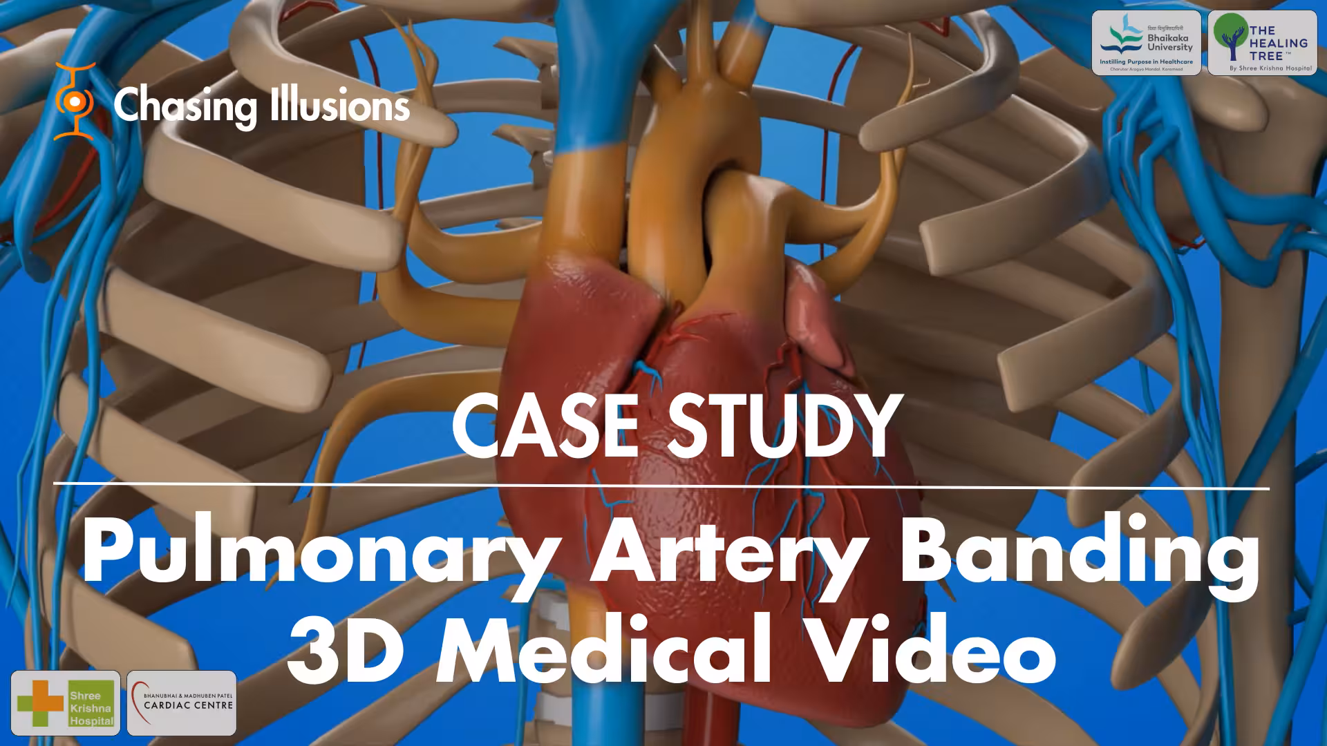
Pulmonary Artery Banding 3D Medical Video Case Study
Introduction: Pulmonary Artery Banding
This case study showcases how our medical animation team created a 3D Pulmonary Artery Banding medical video. We used immersive, engaging visuals and seamless storytelling to explain this critical surgical procedure, originally developed in 1952 to address excessive pulmonary blood flow in pediatric patients. The video ensures absolute medical accuracy and anatomical correctness, making a complex topic accessible to a wider audience.
Client Video: Pulmonary Artery Banding 3D Medical Video
Highlights: Pulmonary Artery Banding 3D Medical Video
Historical Context: First reported by Mueller and Damon in 1952, establishing its importance in pediatric cardiology.
Surgical Approaches: Comparison of left lateral thoracotomy and median sternotomy, with rising preference for median sternotomy.
Technical Process: Detailed methodology of creating and tightening the band around the pulmonary artery using specific tools and sutures.
Ventilation Impact: Median sternotomy offers minimal interference with ventilation, facilitating complex surgeries.
Precision in Measurement: Use of catheters to monitor distal pulmonary artery pressure during the procedure.
Future Procedures: Single incision benefits for subsequent surgeries post median sternotomy approach.
Patient Preparation: Importance of PA banding in managing left ventricle retention for patients with transposition of great arteries.
Key Insights: Pulmonary Artery Banding 3D Medical Video
Evolution of Technique: The historical context of pulmonary artery banding reflects innovations in pediatric surgical practice. Initially, techniques varied, but evolving surgical needs and technological advances have streamlined such procedures, emphasizing the importance of ongoing education and adaptation in surgery.
Decision-Making in Surgery: Choosing between surgical approaches requires a nuanced understanding of individual patient anatomy and pathology. The trend toward median sternotomy underscores the importance of tailoring surgical decisions to improve outcomes and operating efficiency.
Impact of Surgical Access: The clinical advantage of median sternotomy not only facilitates immediate surgical access but also prepares the surgical field for future procedures, showcasing the strategic planning involved in pediatric cardiothoracic surgeries.
Anatomical Considerations: The careful dissection and handling of surrounding structures, such as the aorta and small veins, exemplify the meticulous nature of cardiac surgery and highlight the potential challenges surgeons face during such procedures.
Monitoring Technology: Using catheters for real-time monitoring of pulmonary artery pressure demonstrates the integration of technology in surgical practice. This level of monitoring is crucial for ensuring the safety and efficacy of complex cardiac procedures.
Suture Techniques: The different methodologies for securing the pulmonary artery banding reveal the importance of surgical skills in ensuring the stability of the intervention, which is vital for achieving desired physiological outcomes post-surgery.
Long-Term Patient Management: The discussion on preparing the left ventricle for further surgical interventions indicates an understanding of long-term patient outcomes, showcasing how immediate surgical decisions impact future cardiac health and management strategies.
Client Overview
Name: Dr. Vishal Bhende, Pediatric Cardio Thoracic Surgeon at Bhanubhai & Madhuben Patel Cardiac Centre
Industry: Healthcare
About: Dr. Vishal Bhende, a Pediatric Cardio Thoracic Surgeon at the Bhanubhai & Madhuben Patel Cardiac Centre of Shree Krishna Hospital, is recognized for his innovative approach to medical education. He transforms complex surgical procedures into simple, engaging 3D videos, making them easy to understand for medical professionals, students, and patients alike.
Dr. Vishal V. Bhende
Dr. Bhende’s commitment to advancing medical knowledge is further highlighted by his Guinness World Record for co-authoring a peer-reviewed academic paper with 15,025 contributors from the COVIDSurg and GlobalSurg Collaboratives at the University of Birmingham.
Contribution: Expanding on his innovative approach, Dr. Bhende created a YouTube channel, Dr. Vishal V Bhende, to showcase his animated medical videos. The channel features a wide range of complex procedures, including Eponymous Cardiovascular Surgeries, Atrio-ventricular Canal Repair Techniques, Hydatid Cyst Barrett’s Method, Modified Senning’s Operation, Intra Operative Scimitar Syndrome, etc.,, making them simple and accessible for the medical community.
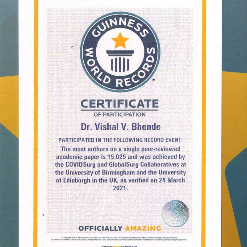
Source: Guinness World Record
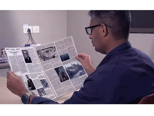
His excellent medical knowledge & success as a surgeon expert led him to publish his article at TimesofIndia (Pediatric Cardio Thoracic Surgeon developed Heart Surgery 3D Animation Video), in which he discussed how he helped the medical industry to join animation technology to bring the revolution in complex medical procedures by converting them into engaging & scientifically visualized medical videos.
Why Choose Us
Dr. Vishal Bhende chose Chasing Illusions for our expertise in creating visually engaging and medically accurate 3D surgical animations. Our 3D medical explainer videos simplifies the complex procedure for addressing excessive pulmonary blood flow, highlighting critical cardiac structures and procedural steps with exceptional detail for professionals and patients.
By this, Dr. Vishal Bhende can simplify the complex subject of congenital heart disease, further, he can enhance medical presentations, boost surgeon training programs, and improve patient understanding in a compelling and easily digestible format.
Client Challenge
The healthcare industry is experiencing a significant transformation, with medical professionals eagerly adopting new technologies. This shift is revolutionizing medical education by changing how concepts are taught and understood. Among these innovations, 3D medical animation has emerged as a key tool, profoundly enriching the learning experience.
Faced with the challenge of accurately representing the complex Pulmonary Artery Banding procedure, Dr. Vishal Bhende of the Bhanubhai & Madhuben Patel Cardiac Centre needed a medical animation agency who could create a detailed and engaging 3D medical animation. The goal was to find an excellent 3D medical video maker who could deliver high-quality work within a specific budget and timeframe.
Apart from this, he wanted to educate the medical practitioners and professional surgeons effectively in simplifying the complex subject of congenital heart disease and its treatment by ensuring absolute medical accuracy and anatomical correctness throughout the whole process.
Our Process

Credit: Chasing Illusions
Chirag Anand, the founder of Chasing Illusions, began by working closely with Dr. Vishal Bhende and his medical team. Together, they conducted in-depth research to ensure complete accuracy. Their shared goal was to create a 3D medical animation that could help a wide audience—from experienced cardiac surgeons to medical students and patients—easily understand a complex cardiac surgical procedure.
After the initial research, we developed a detailed storyboard to visually map out each critical step of the procedure. Once Dr. Bhende approved it, our 3D medical aniamators brought the concept to life. They used realistic modeling, texturing, and animation to highlight every anatomical and surgical detail with exceptional clarity.
Throughout the process, we maintain close communication with the doctors’ team, especially Dr. Vishal, ensuring the final 3d surgical medical video is both scientifically precise and visually engaging for education, training, or patient communication.
Solution

Source: Chasing Illusions
The final result was a 3-minute and 33-second 3D medical video that met all of Dr. Bhende’s needs. By combining scientific accuracy with compelling visuals, the animation transforms the complex Pulmonary Artery Banding procedure into an easily digestible format, complete with unmatched anatomical and surgical detail.
This 3D Pulmonary Artery Banding video was designed to provide medical professionals with a clear understanding of the challenges associated with excessive pulmonary blood flow and how to surgically address them. The project also serves as a testament to the power of science to solve critical medical problems through precise and impactful educational tools.
Results
Dr. Vishal Bhende, Pediatric Cardio Thoracic Surgeon at Bhanubhai & Madhuben Patel Cardiac Centre of Shree Krishna Hospital’s decision to trust in Chasing Illusions for Pulmonary Artery Banding 3D Medical Video brought significant results:
1. First, the video helped to enhance surgical training in which surgeons and medical staff gained a clear, visual understanding of each step in the procedure, improving training effectiveness.
2. Second, the patients and their families could easily understand the complex medical procedures, leading to increased trust and more informed consent.
3. Third, the hospital increased its educational resources for workshops, seminars, and internal training programs that invited new medical trainees could comprehend complex anatomical and procedural details faster through high-quality visual aids.
4. Fourth, the video served as a powerful knowledge-sharing tool for cross learning platforms, which helped Shree Krishna Hospital to show its commitment to advanced education tools, strengthening its reputation for medical excellence.
The goal was to create an engaging medical visual story that would educate, engage, and convert.
Summary: Pulmonary Artery Banding 3D Medical Video
First reported in 1952, Pulmonary Artery Banding (PAB) is a surgical procedure for children with excessive pulmonary blood flow from large shunts or single ventricle anomalies. Traditionally performed via a left lateral thoracotomy, the procedure is now more commonly done through a median sternotomy. This approach offers several advantages, including better access to complex vascular structures and easier follow-up surgeries, all through a single incision.

Source: Chasing Illusions
The surgical technique for Pulmonary Artery Banding involves carefully dissecting the area between the aorta and the main pulmonary artery. A strip of PTFE membrane is then placed around the pulmonary artery to create a band, which is gradually tightened to restrict blood flow. The pressure is carefully monitored to ensure the correct degree of constriction. This procedure not only addresses excessive pulmonary outflow but also prepares the left ventricle for corrective surgery in cases of transposition of the great arteries.
Conclusion: Pulmonary Artery Banding
The 3D medical video for Pulmonary Artery Banding, created for Dr. Vishal Bhende by Chasing Illusions and founder Chirag Anand, has become a powerful tool. It has proven effective in advancing surgical education, enhancing patient communication, and supporting the growth of medical institutions.
By converting complex medical procedures into clear and engaging visuals, our medical animation videos served as a valuable asset for patient education, surgical team preparation, and showcasing the hospital’s specialized expertise.
By focusing on accuracy, clarity, and close collaboration, we exceeded Dr. Bhende’s expectations. The final video positions the Bhanubhai & Madhuben Patel Cardiac Centre at the forefront of modern medical education and communication.
CONTACT NOW
HURRY UP!!! Your Medical Video is Just One Step Away! Right Now WhatsApp Us