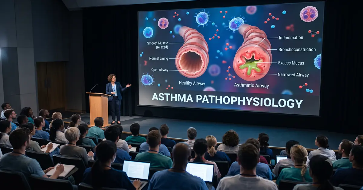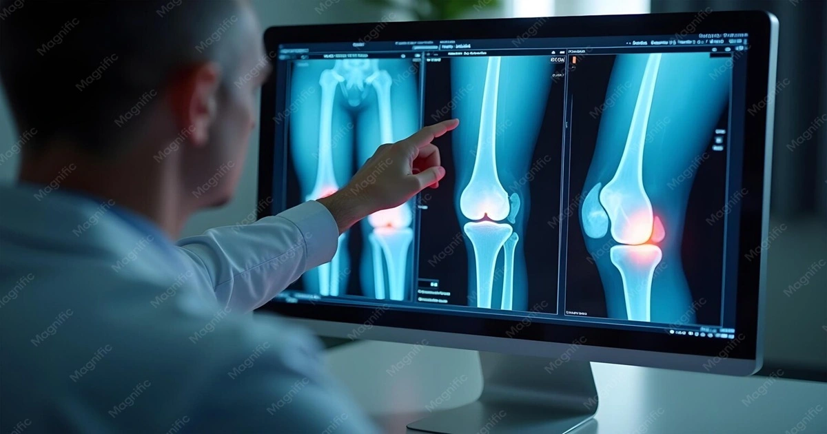
Imagine being told that your damaged knee joint will be reshaped, diseased cartilage will be removed, and several precisely engineered components will be positioned inside the joint. For a patient, medical student, or even a healthcare professional learning a new implant system, understanding all of that from text and static illustrations alone can be difficult.
This is where Total Knee Replacement 3D Medical Animation becomes especially valuable. Instead of asking the viewer to mentally reconstruct a complex surgical procedure, 3D medical visualization can show the knee anatomy, damaged joint surfaces, bone preparation, implant positioning, and reconstructed joint as a continuous visual sequence.
The objective is not to replace the orthopedic surgeon's explanation, clinical training, or surgical guidance. It is to make complex information easier to see, understand, discuss, and remember.
According to the U.S. National Library of Medicine's MedlinePlus, knee replacement surgery involves removing damaged cartilage and some bone and replacing the affected joint surfaces with artificial components. Total knee replacement generally addresses the surfaces at the end of the femur, the top of the tibia, and, in some procedures, the back of the patella.
Why Is Total Knee Replacement Difficult to Explain with Text Alone?
A total knee replacement, also called total knee arthroplasty (TKA), involves three-dimensional anatomy, multiple surgical stages, precise bone preparation, and the placement of prosthetic components.
A written explanation might say:
Damaged surfaces of the femur and tibia are removed and replaced with prosthetic components.
Clinically, that statement is accurate—but it leaves several questions unanswered for a learner:
Which surfaces are damaged?
Where exactly is cartilage removed?
How much bone is prepared?
How is the femoral component oriented?
Where does the tibial component sit?
What separates the metallic components?
What happens to the patella?
How does the reconstructed knee move after implantation?
These are fundamentally spatial and sequential questions.
A 3D medical animation can answer them visually by moving from an intact knee to an arthritic joint, then progressively revealing each surgical stage.
That is why surgical animation is particularly useful for orthopedic education: the learner does not merely read what happens; they can see where, when, and in what sequence it happens.
Understanding the Knee Before Understanding Knee Replacement
Before demonstrating surgery, an effective medical animation should establish the anatomy of the healthy knee.
The knee is primarily formed by:
Femur: the thigh bone
Tibia: the larger lower-leg bone
Patella: the kneecap
Articular cartilage: smooth tissue covering the ends of bones
Menisci: fibrocartilage structures that contribute to load distribution
Ligaments: structures that help stabilize the knee
Joint capsule and surrounding soft tissues
Healthy articular cartilage provides a smooth surface that helps the bones move relative to one another.
In osteoarthritis, this cartilage can progressively deteriorate. As the joint surfaces become damaged, movement may become painful and stiff. Osteoarthritis is the most common condition leading to knee replacement surgery.
How 3D Animation Makes the Difference?
Instead of placing healthy and arthritic knees in two separate textbook images, an animation can transition between them.
For example:
Healthy joint → cartilage degeneration → narrowing of joint space → irregular joint surfaces → advanced arthritic knee
The viewer can immediately understand what has changed anatomically and why those changes can interfere with joint movement.
This establishes the clinical context before showing the surgery itself.
How Total Knee Replacement 3D Medical Animation Explains the Procedure?
An effective Total Knee Replacement 3D Medical Animation should not simply show an implant appearing inside the knee. It should tell a logical visual story.
The procedure can be explained through the following stages.
Stage 1: Showing the Damaged Knee Joint
The animation can begin with an anatomically accurate external view of the knee before transitioning beneath the skin to the bones and joint surfaces.
The camera can then focus on areas of:
damaged cartilage,
degenerative joint surfaces,
reduced joint space,
exposed or irregular bone surfaces,
osteophytes when relevant, and
altered joint mechanics.
A transparent or cutaway visualization can make structures visible without requiring the viewer to interpret complicated overlapping anatomy.
Why This Matters?
Patients may know that they have "arthritis," but that term does not necessarily help them visualize what has physically changed inside their knee.
For medical learners, the visualization creates a bridge between pathology and the surgical intervention that follows.
Stage 2: Visualizing Surgical Access to the Knee
The next sequence can demonstrate the surgical approach in an educational, non-graphic manner.
Rather than focusing on blood or tissue trauma, the visualization can simplify the surgical field and show how the surgeon gains access to the joint.
For example, the animation can:
isolate the knee anatomically,
reveal deeper structures through controlled transparency,
show the patella being repositioned where appropriate,
expose the femoral and tibial joint surfaces.
The NHS describes total knee replacement as involving access to the knee joint followed by removal of the damaged ends of the thigh bone and shin bone before the replacement components are fitted.
For patient education, this approach keeps the visualization clinically informative without making it unnecessarily graphic.
Stage 3: Removal of Damaged Joint Surfaces
This is one of the most important sequences in a knee replacement surgery animation.
The viewer should be able to distinguish between:
Before: damaged articular surfaces
and
After: prepared bone surfaces ready to receive the prosthetic components.
MedlinePlus explains that damaged cartilage and some bone are removed before artificial components are attached to the prepared surfaces.
What Animation Can Show Clearly?
A well-designed visualization can demonstrate:
the damaged distal femoral surface,
the affected proximal tibial surface,
the transition from diseased cartilage to prepared bone,
the relationship between the bone cuts and future implant placement.
Different colors or carefully controlled material properties can visually distinguish:
cartilage,
cortical and cancellous bone,
surgical instruments,
trial components,
final prosthetic components.
This is substantially easier to interpret than describing every spatial relationship in text.
Stage 4: Femoral Bone Preparation
The distal femur must be prepared so the femoral component can fit appropriately.
This is a particularly useful stage for 3D knee replacement animation because the femur has complex three-dimensional geometry.
An educational animation can rotate around the joint and demonstrate how the femoral surface changes from its natural contour to the prepared geometry required for the implant.
Useful Visual Techniques
Medical animators can use:
transparent overlays,
before-and-after comparisons,
ghosted implant positioning,
isolated anatomical views,
cross-sectional views,
controlled camera rotation.
For a medical student or device-training audience, these techniques can make implant orientation considerably easier to comprehend.
Stage 5: Tibial Bone Preparation
The upper surface of the tibia is also prepared to receive the tibial component.
A knee surgery medical animation can clearly show the relationship between:
proximal tibia,
prepared tibial surface,
tibial tray,
polyethylene insert,
femoral component.
A static illustration may show these components individually. Animation can demonstrate how they fit together dynamically.
This becomes especially useful when teaching the difference between anatomical bone surfaces and the engineered surfaces of an artificial knee joint.
Stage 6: Understanding the Prosthetic Knee Components
The prosthesis is not simply one artificial "knee."
It generally consists of multiple components working together.
Femoral Component
The femoral component covers the prepared end of the femur.
Tibial Component
A tibial baseplate or tray is positioned on the prepared upper surface of the tibia.
Polyethylene Insert
A durable polymer bearing surface is typically positioned between the femoral and tibial components, allowing the articulating surfaces to move relative to each other.
Patellar Component
In some total knee replacement procedures, the surface on the back of the patella may also be resurfaced with a prosthetic component.
MedlinePlus describes knee prostheses as involving artificial components positioned at the lower femur, upper tibia, and potentially the back of the kneecap.
Why an Exploded 3D View Works So Well?
This stage is ideal for an exploded-view animation.
The components can separate from the knee, rotate individually, and then return to their anatomical positions.
For orthopedic implant manufacturers, healthcare educators, and clinical training teams, an exploded visualization can communicate:
implant geometry,
component relationships,
positioning,
articulation,
system configuration.
Complex engineering and anatomy can therefore be communicated in the same visual environment.
Stage 7: Trial Positioning and Alignment
Simply placing an implant inside a generic knee model does not adequately explain knee arthroplasty.
Positioning matters.
An advanced Total Knee Replacement 3D Medical Animation can show how trial or final components relate to:
femoral anatomy,
tibial anatomy,
joint alignment,
joint spacing,
surrounding structures.
For professional audiences, visualization can also be adapted to explain specific surgical techniques, implant systems, instrumentation, or technology-assisted workflows.
The animation must remain aligned with the specific procedure being taught rather than implying that every total knee replacement is performed identically.
Stage 8: Implant Placement
Once the bone surfaces have been prepared, the visualization can show the final components moving into position.
The sequence can demonstrate:
Femoral component → tibial component → bearing surface → optional patellar component → reconstructed knee
The NHS describes the replacement surfaces as commonly involving metal and plastic components positioned over the prepared ends of the femur and tibia.
The FDA also recognizes specific safety and performance requirements for knee-joint replacement implants through standards addressing factors including design, materials, evaluation, manufacture, sterilization, packaging, and testing.
For medical device communication, this is important: visualization should accurately represent the actual implant rather than substitute a generic prosthesis when product-specific education is intended.
Stage 9: Showing the Reconstructed Knee in Motion
One of the greatest advantages of animation comes after the components have been positioned.
A static rendering can show the final implant.
Animation can show what the reconstructed joint is designed to do.
The knee can be animated through controlled flexion and extension while the camera shows how the prosthetic surfaces articulate.
A useful sequence might show:
Damaged knee motion → surgical reconstruction → prosthetic joint motion
This closes the visual learning loop.
The audience sees:
what was damaged,
what was removed,
what replaced it,
where the components were positioned,
how the reconstructed joint relates mechanically.
CASE STUDY: Total Knee Replacement Surgery Video
Why Medical Animation Works Especially Well for Orthopedic Surgery?
Orthopedic surgery combines anatomy, biomechanics, surgical technique, and medical-device engineering.
That makes it particularly suitable for 3D visualization.
1. It Reveals Anatomy Hidden Inside the Body
The camera can move beneath the skin without disrupting anatomical orientation.
Bones, cartilage, ligaments, implants, and surgical instruments can be selectively displayed or hidden.
2. It Shows the Procedure as a Sequence
Total knee replacement is not a single event.
Animation can visually connect:
Disease → surgical access → bone preparation → component positioning → reconstruction
That sequencing can reduce the cognitive effort required to mentally assemble information from separate diagrams.
3. It Allows Impossible Camera Angles
A real surgical camera is constrained by the operating field.
A virtual 3D camera is not.
It can move:
inside the joint,
underneath a prosthetic component,
around the posterior side of the knee,
through transparent tissue,
directly along an implant axis.
Those perspectives can be valuable for explaining spatial relationships.
4. It Can Control Complexity
Not every audience needs the same amount of information.
A patient may require a simplified anatomical explanation.
An orthopedic resident may require considerably more surgical detail.
A device representative may need an emphasis on implant design.
The same core 3D assets can therefore support multiple educational levels when the content is appropriately designed and medically reviewed.
3D Medical Animation vs Text and Static Images
Medical textbooks and anatomical illustrations remain essential educational tools. Medical animation should complement them rather than be presented as a replacement.
However, the formats solve different communication problems.
Educational Need | Text | Static Illustration | 3D Medical Animation |
|---|---|---|---|
Define terminology | Excellent | Moderate | Good |
Show knee anatomy | Moderate | Excellent | Excellent |
Demonstrate depth | Limited | Moderate | Excellent |
Explain surgical sequence | Moderate | Limited | Excellent |
Show implant positioning | Limited | Good | Excellent |
Demonstrate joint movement | Limited | Limited | Excellent |
Repeat a procedure visually | Limited | Limited | Excellent |
Show hidden structures selectively | Difficult | Moderate | Excellent |
Compare pre- and post-surgical anatomy | Good | Good | Excellent |
The strongest medical education often combines these formats rather than relying exclusively on one.
How 3D Medical Animation Supports Patient Education?
Patients approaching knee replacement can encounter unfamiliar terms such as:
arthroplasty,
femoral component,
tibial component,
polyethylene insert,
resurfacing,
prosthesis,
joint alignment.
Explaining each term verbally during a consultation can become information-heavy.
A knee replacement procedure video can establish a shared visual reference between the clinician and patient.
Rather than merely saying, "The damaged surface will be removed," a physician can point to the exact area being discussed.
Animation Can Help Explain
where the knee damage is located,
what joint replacement means,
which surfaces are prepared,
where the implants are positioned,
how the artificial surfaces interact,
the difference between total and partial knee replacement,
the general purpose of rehabilitation after surgery.
MedlinePlus specifically provides patient-oriented knee replacement educational resources and emphasizes discussing the risks, benefits, and suitability of surgery with a healthcare professional.
Important Limitation
A medical animation should never imply that every patient will experience the same surgical technique, recovery timeline, outcome, or implant selection.
Patient-specific decisions belong to the treating orthopedic team.
How Total Knee Replacement Animation Supports Medical Students?
Medical students must connect anatomy with surgical intervention.
A textbook may explain the femur, tibia, patella, articular cartilage, and ligaments separately.
A total knee arthroplasty animation can bring those structures together in a clinical scenario.
Students can observe:
normal anatomy,
osteoarthritic changes,
surgical exposure,
bone preparation,
component placement,
reconstructed joint anatomy.
This progression supports a more integrated understanding of the procedure.
For healthcare eLearning, the animation can also be divided into modular chapters, allowing students to replay individual surgical stages.
How 3D Animation Supports Orthopedic Surgical Education?
For surgeons, residents, fellows, and clinical training teams, the required level of detail is substantially higher.
A professional orthopedic medical animation may include:
detailed bone morphology,
surgical planes,
implant geometry,
instrumentation,
component sizing concepts,
trial components,
orientation,
fixation,
articulated postoperative views.
The objective is not to teach surgery through animation alone. Rather, animation can supplement established surgical training, supervised clinical education, cadaveric learning, simulation, surgical documentation, and manufacturer-specific training.
When the intended audience is professional, medical accuracy and review by subject-matter experts become especially important.
How Knee Implant Companies Can Use 3D Medical Animation?
Orthopedic implants are difficult products to communicate through conventional marketing imagery because much of their function occurs inside the body.
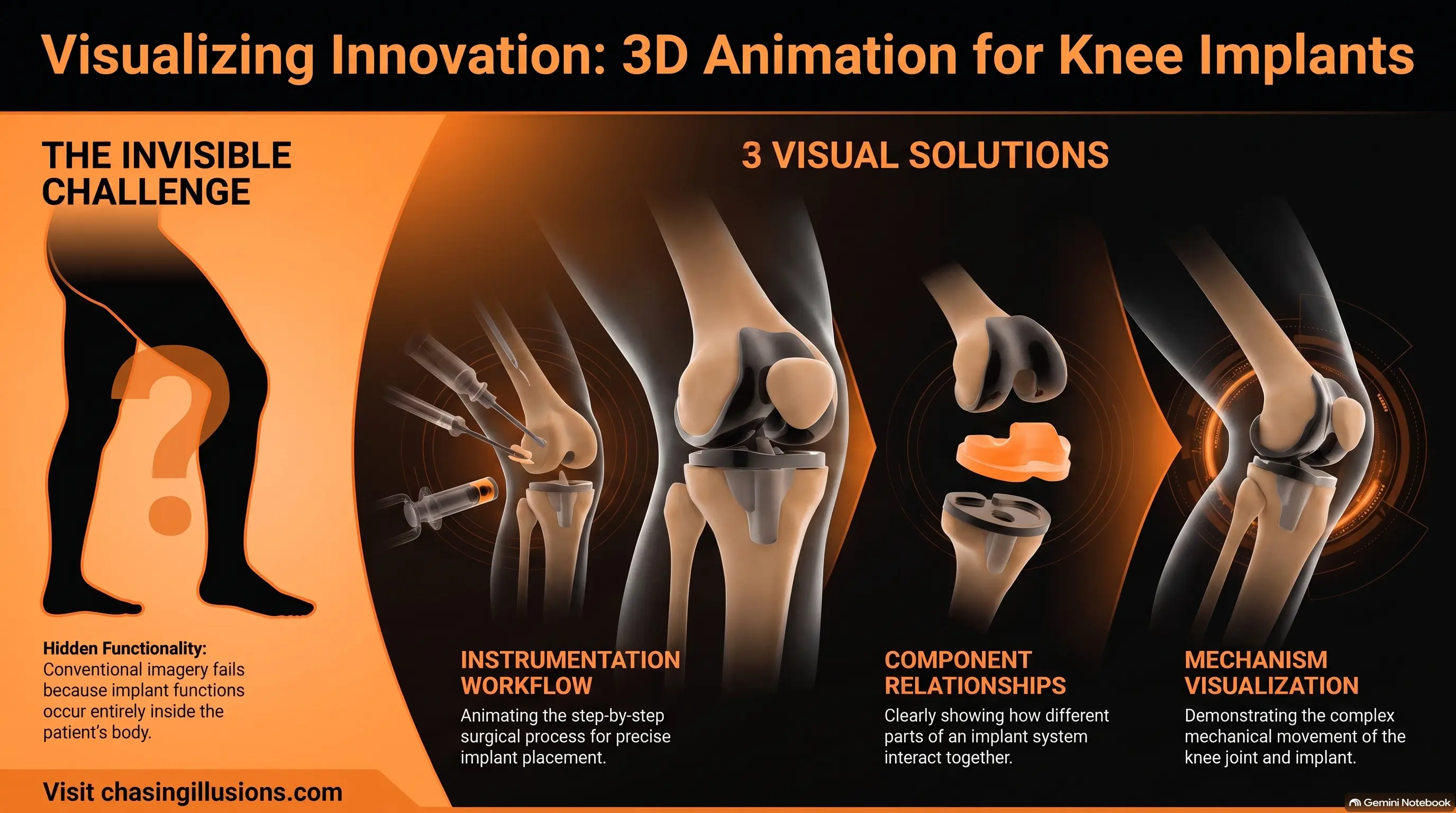
A knee implant animation can demonstrate both the engineering and clinical context.
Implant System Visualization
Individual components can be isolated, rotated, sectioned, and examined.
Instrumentation Workflow
Product-specific instruments can be shown interacting with accurate anatomical models.
Component Relationships
Exploded views can explain how the femoral component, tibial baseplate, insert, and patellar component relate spatially.
Implant Positioning
The implant can be shown entering its final anatomical position without the visual obstruction present in real surgical footage.
Mechanism Visualization
Animation can demonstrate articulation after reconstruction.
Because implant design and approved indications differ between systems, device-specific visualizations should be based on verified product data and reviewed for regulatory and clinical accuracy.
Explore our 3D Medical Animations Portfolio for Healthcare Professionals
Why Anatomical Accuracy Matters in Knee Replacement Animation?
Beautiful animation is not automatically good medical animation.
A clinically useful 3D medical animation for knee surgery requires accurate representation of:
bone anatomy,
cartilage,
implant geometry,
component relationships,
surgical sequence,
instrument interaction,
anatomical orientation,
movement.
Small inaccuracies can create major educational misconceptions.
For example, an implant positioned incorrectly by only a visually subtle amount may still look acceptable to a general audience while being misleading to an orthopedic specialist.
This is why professional medical-animation workflows generally benefit from collaboration among:
medical animators,
orthopedic subject-matter experts,
surgeons,
medical writers,
device engineers where applicable,
scientific reviewers.
For projects involving orthopedic procedures and implant communication, Chasing Illusions Studio can develop medically oriented 3D visualizations around approved scripts, anatomical references, device CAD data, and subject-matter-expert feedback.
What Makes an Effective Total Knee Replacement Surgery Video?
A high-quality medical explainer should prioritize comprehension over visual spectacle.
Accurate Anatomy
The femur, tibia, patella, articular surfaces, and relevant surrounding structures should maintain realistic spatial relationships.
Clear Visual Hierarchy
The viewer should always know what they are supposed to observe.
Unnecessary anatomical detail can distract from the instructional objective.
Controlled Camera Movement
Camera movement should clarify anatomy—not simply make the video feel cinematic.
Step-by-Step Progression
The procedure should follow a logical educational sequence.
Consistent Color Coding
If cartilage, bone, implants, or instruments are differentiated through color, the visual language should remain consistent.
Appropriate Transparency
Transparency can expose otherwise hidden structures while preserving anatomical orientation.
Audience-Specific Detail
Patient education and orthopedic surgical education should not use exactly the same script or visual complexity.
Medical Review
Final animation should be reviewed for scientific and clinical accuracy before publication.
A Practical Visual Storyboard for Total Knee Replacement
A complete educational sequence could follow this structure:
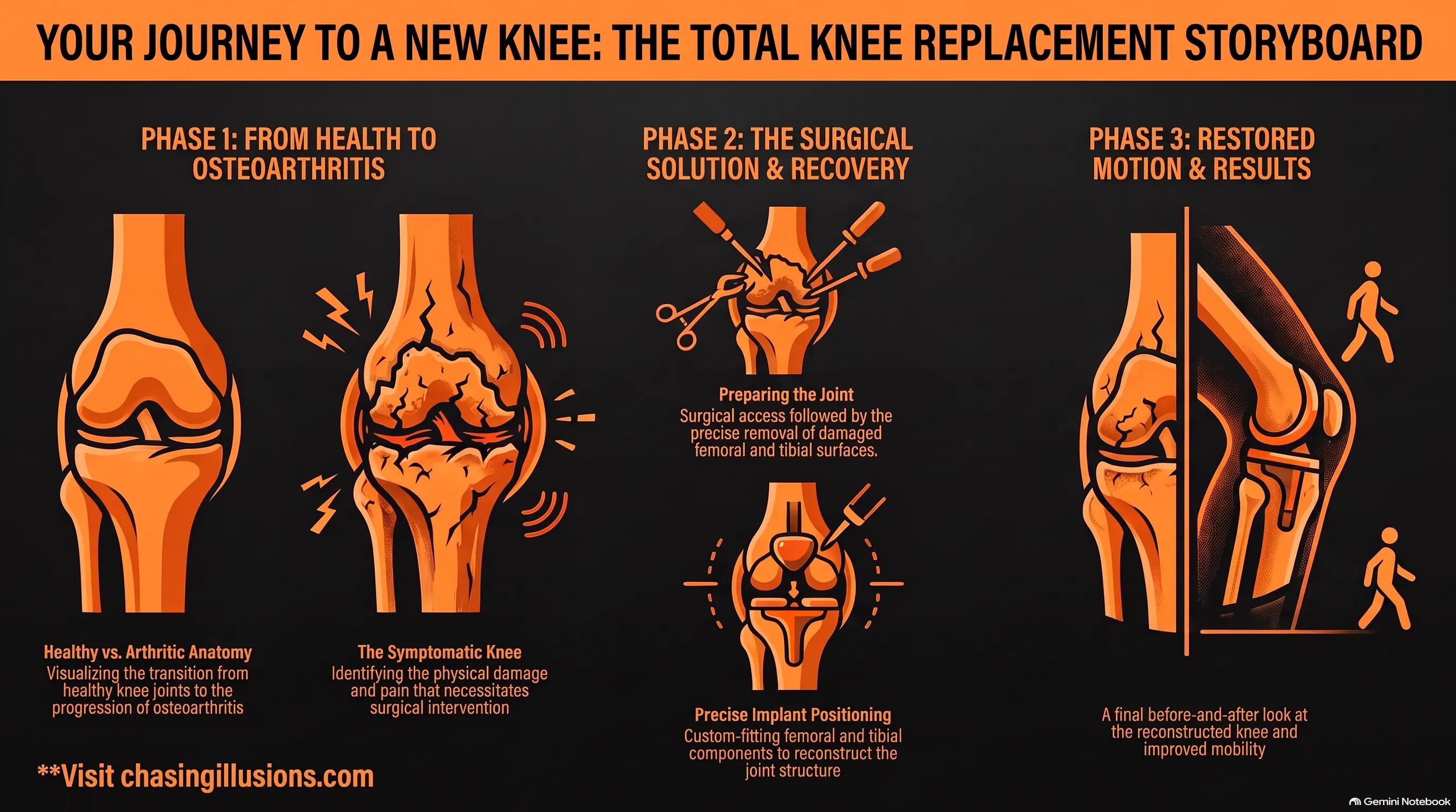
Scene 1 — Healthy Knee Anatomy
Show the femur, tibia, patella, and smooth joint surfaces.
Scene 2 — Osteoarthritis Progression
Visualize damaged cartilage and deteriorating joint surfaces.
Scene 3 — Symptomatic Arthritic Knee
Demonstrate why damaged surfaces can interfere with comfortable movement.
Scene 4 — Surgical Access
Reveal the knee joint using a clean, non-graphic surgical visualization.
Scene 5 — Damaged Surface Removal
Show preparation of the femoral and tibial surfaces.
Scene 6 — Femoral Preparation
Highlight the prepared distal femur.
Scene 7 — Tibial Preparation
Show the prepared proximal tibia.
Scene 8 — Implant Components
Introduce the femoral, tibial, polyethylene, and applicable patellar components using an exploded 3D view.
Scene 9 — Implant Positioning
Demonstrate each component moving into its anatomical position.
Scene 10 — Final Reconstruction
Show the completed artificial knee joint.
Scene 11 — Motion
Demonstrate controlled flexion and extension.
Scene 12 — Before-and-After Comparison
Compare the damaged knee anatomy with the reconstructed joint.
This creates a coherent narrative instead of presenting disconnected surgical facts.
Total Knee Replacement vs Partial Knee Replacement: Animation Can Clarify the Difference
These procedures are sometimes confused by patients.
A total knee replacement addresses multiple joint surfaces, whereas partial knee replacement replaces only the affected portion of the joint when clinically appropriate. MedlinePlus notes that partial knee replacement is intended for damage primarily involving one portion of the knee, while total replacement addresses the whole joint.
A split-screen animation can make the difference immediately visible:
Partial replacement: preserved healthy compartments remain visible.
Total replacement: broader femoral and tibial joint surfaces are reconstructed.
This can often communicate the distinction faster than several paragraphs of description.
Can 3D Animation Replace a Surgeon's Explanation?
No.
Total Knee Replacement 3D Medical Animation is an educational communication tool—not an alternative to medical consultation, informed consent, clinical judgment, or supervised surgical training.
Its value is in supporting those processes.
For patients, it may help them formulate better questions.
For students, it can provide visual context.
For healthcare educators, it offers a repeatable teaching resource.
For implant manufacturers, it can communicate complex device functionality.
For clinicians, it can provide another way to explain anatomy and procedural concepts.
Frequently Asked Questions:
What is total knee replacement?
Total knee replacement, or total knee arthroplasty, is a surgical procedure in which damaged knee joint surfaces are removed or prepared and replaced with artificial components. It is commonly performed for severe joint damage associated with conditions such as osteoarthritis when appropriate non-surgical treatment has not provided adequate relief.
What parts are replaced during total knee replacement?
The procedure generally involves the distal femur and proximal tibia. Depending on the surgical approach and patient circumstances, the back of the patella may also be resurfaced.
What are knee replacement implants made from?
Knee prostheses commonly use metallic components together with durable polymer bearing materials. Exact materials and designs vary by implant system. FDA-recognized standards for knee-joint replacement implants address metallic and non-metallic components and requirements concerning design, performance, manufacture, sterilization, and testing.
Why use 3D animation to explain knee replacement?
The procedure involves anatomy, depth, movement, bone preparation, multiple implant components, and a sequence of surgical actions. 3D animation allows these relationships to be visualized dynamically rather than forcing the learner to reconstruct the procedure mentally from text.
Is a total knee replacement animation suitable for patients?
Yes, provided the level of anatomical and surgical detail is adapted for patient education and the animation clearly states that individual procedures and outcomes differ.
Can medical students use knee replacement animations?
Yes. Animation can supplement anatomy teaching, orthopedic education, medical eLearning, lectures, simulation, and other established learning methods.
Can implant manufacturers use customized animations?
Yes. Product-specific animation can demonstrate implant components, device geometry, instrumentation, assembly relationships, or procedural concepts when created from validated technical and clinical information.
Does medical animation replace surgical training?
No. Surgical animation is a supplementary educational resource. It cannot replace supervised clinical training, formal surgical education, hands-on experience, or professional medical judgment.
Conclusion: Making an Invisible Surgical Journey Visible
Total knee replacement becomes difficult to understand when the learner must imagine an entire three-dimensional surgical sequence from words and isolated images.
3D medical animation changes the educational perspective.
Instead of simply telling the audience that damaged bone and cartilage are removed and prosthetic components are inserted, it can visually connect:
damaged joint → surgical preparation → bone resurfacing → implant positioning → reconstructed knee → joint movement
That continuity is what makes Total Knee Replacement 3D Medical Animation particularly valuable for patient education, orthopedic education, medical schools, clinical training, healthcare eLearning, and orthopedic device communication.
Medical animation does not simplify the science by removing important information. When produced correctly, it simplifies access to the science by making anatomy, spatial relationships, surgical progression, and device positioning visible.
Need to Explain a Complex Orthopedic Procedure Visually?
If your hospital, orthopedic team, healthcare education platform, medical school, or implant company needs to communicate a complex knee procedure more clearly, Chasing Illusions Studio can create custom 3D medical animation and healthcare explainer videos based on your educational objectives, clinical references, and approved technical materials.
Turn complex orthopedic information into a visual learning experience your audience can understand.
NOTE: This article is intended for general educational and medical-communication purposes. It does not provide individual medical advice, diagnosis, treatment recommendations, or surgical instruction. Patients should discuss their condition, implant options, risks, benefits, and expected recovery with a qualified orthopedic healthcare professional.
Book a discovery 20-minutes strategy call.
📞 +91-9910911696 | +91-9910660851
📩 info [@] chasingillusions [.] in
Written by Deepak, Healthcare Content Strategist at Chasing Illusions Studio, who leads content for clients including Ambler Surgical, Practo, Bayer, SMT, Novartis, and 100+ healthcare brands across India, USA, Thailand, and the UK.
Last Updated: Aug 26 2026 | Chasing Illusions Studio
Chasing Illusions Studio
Premium animation & video production studio based in Delhi, India. Specialising in 3D animation, medical visualisation, architectural walkthroughs, and CGI.