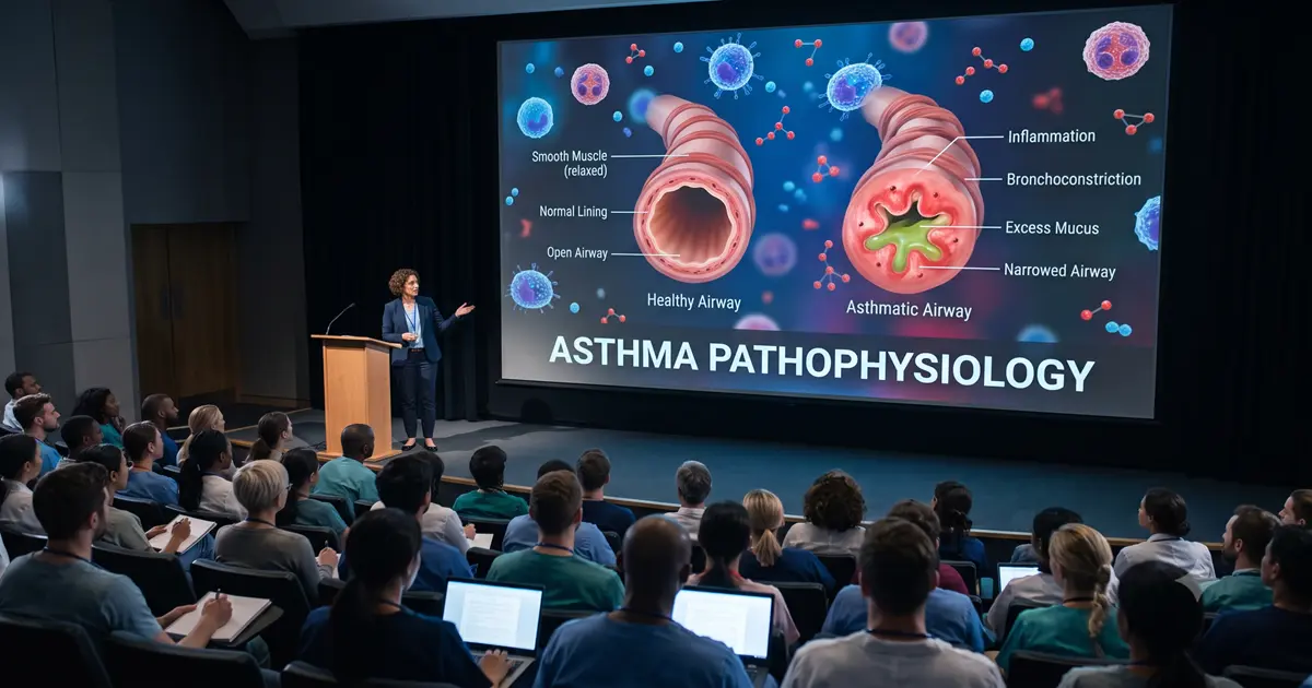
Imagine trying to explain why a patient suddenly struggles to breathe using only a flat diagram of the lungs. The diagram may identify the bronchial tubes, but it cannot fully show the airway wall swelling, smooth muscles tightening, mucus accumulating, and airflow becoming restricted in real time.
A 3D Medical Animation of Asthma Pathophysiology transforms these invisible biological processes into a clear visual sequence. It helps medical students, clinicians, respiratory therapists, pharmaceutical teams, and patients understand what happens inside the lungs before, during, and after an asthma episode.
What Is Asthma Pathophysiology?
Asthma is a heterogeneous chronic respiratory disease characterized by variable respiratory symptoms, airway inflammation, bronchial hyperresponsiveness, and fluctuating expiratory airflow limitation. Its manifestations can differ substantially between individuals because multiple inflammatory pathways, triggers, phenotypes, and endotypes may be involved.
According to the Global Initiative for Asthma, asthma is a heterogeneous chronic respiratory disease characterized by variable symptoms, airway inflammation, bronchial hyperresponsiveness, and variable expiratory airflow limitation.
The World Health Organization describes asthma as a chronic lung disease in which inflammation and muscle tightening around the airways make breathing difficult. Common symptoms include wheezing, coughing, chest tightness, and shortness of breath.
At the physiological level, asthma commonly involves four interconnected changes:
Inflammation and swelling of the airway lining
Contraction of bronchial smooth muscle
Increased mucus production
Heightened airway sensitivity to triggers
The National Heart, Lung, and Blood Institute explains that sensitive airways may swell, narrow, produce additional mucus, and become surrounded by tightened muscles. With time, airway walls may also become thicker.
These processes are dynamic rather than static. That is why animated medical visualization is particularly valuable for explaining the disease.
How a 3D Medical Animation of Asthma Pathophysiology Works?
A scientifically developed asthma animation usually begins with normal respiratory anatomy before gradually revealing the changes associated with an asthmatic response.
The viewer may first travel through the trachea into the bronchi and bronchioles. A cross-sectional view can then compare a healthy airway with an inflamed asthmatic airway.
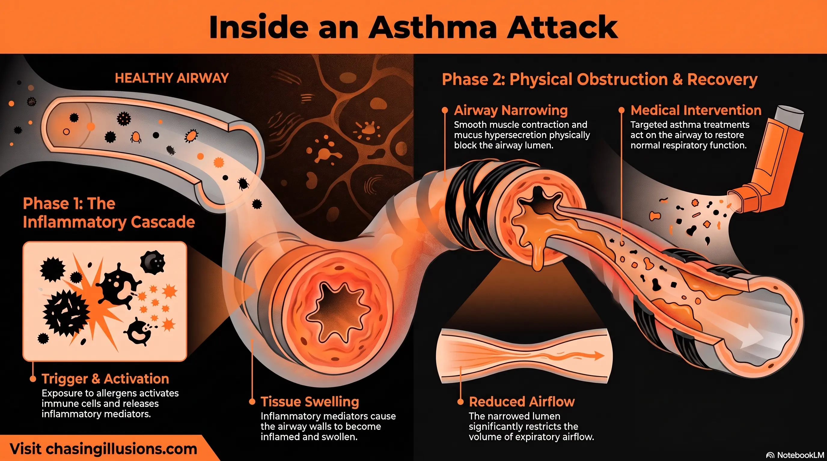
The sequence typically visualizes:
The entry of air into healthy lungs
Exposure to an allergen or another asthma trigger
Activation of epithelial and immune cells
Release of inflammatory mediators
Swelling of the airway wall
Smooth muscle contraction
Mucus hypersecretion
Narrowing of the airway lumen
Reduction in expiratory airflow
The action of relevant asthma treatments
By combining anatomical accuracy with motion, depth, scale, transparency, and controlled camera movement, the animation connects molecular events with the symptoms experienced by a patient.
The Asthma Mechanism Explained Step by Step
1. Exposure to an Asthma Trigger
An asthma episode may be associated with allergens, viral respiratory infections, smoke, air pollution, exercise, occupational exposures, weather changes, or other individual triggers.
In allergic asthma, substances such as pollen, dust mites, animal dander, or mould may interact with the airway epithelium and initiate an immune response. Not every patient responds to the same trigger, and not all asthma is allergic.
A respiratory system animation can demonstrate trigger particles entering the airway and contacting the epithelial surface. This gives learners a clear starting point for the disease mechanism.
2. Airway Epithelial Activation
The airway epithelium is not simply a passive protective layer. When exposed to allergens, pollutants, viruses, or irritants, epithelial cells can release signalling molecules that help initiate and amplify inflammation.
These epithelial-derived signals may include thymic stromal lymphopoietin, IL-25, and IL-33. They can activate immune pathways involving dendritic cells, T-helper type 2 cells, and type 2 innate lymphoid cells.
A detailed asthma pathophysiology animation can magnify the bronchial lining and show epithelial cells releasing signals into surrounding tissue. Colour-coded labels can distinguish airway structures, immune cells, receptors, and inflammatory mediators.
3. Immune Response and Inflammatory Mediators
In many forms of allergic and eosinophilic asthma, type 2 inflammation has an important role. Dendritic cells process allergens and support the activation of T-helper type 2 cells. Cytokines such as IL-4, IL-5, and IL-13 contribute to IgE production, eosinophil recruitment, mucus production, and other inflammatory effects.
Mast cells may release histamine, prostaglandins, leukotrienes, and additional mediators after activation. These substances can promote bronchoconstriction, vascular changes, mucus secretion, and airway inflammation.
Research on eosinophilic asthma shows that eosinophils can release cytokines and granule proteins that contribute to inflammation, epithelial injury, mucus hypersecretion, and airway remodelling.
Medical animation can simplify this complex immune response by presenting it as a logical sequence rather than showing every pathway simultaneously.
4. Eosinophilic Inflammation
Eosinophils are white blood cells involved in many—but not all—asthma phenotypes. IL-5 supports eosinophil development, recruitment, activation, and survival.
Once eosinophils enter airway tissue, they may release inflammatory molecules and cytotoxic granule proteins. These substances can damage the airway epithelium and help sustain inflammation.
However, asthma is biologically diverse. Some patients have eosinophilic or type 2-high inflammation, while others may have neutrophilic, mixed, or paucigranulocytic patterns. Eosinophilic inflammation should therefore not be presented as the universal mechanism in every patient.
An evidence-based pulmonary medical animation should clearly identify the pathway being portrayed—for example, allergic eosinophilic asthma—rather than implying that one immune mechanism represents all asthma.
5. Airway Inflammation and Oedema
Inflammation can make the airway lining swollen and thicker. Increased vascular permeability may contribute to tissue oedema, reducing the diameter of the airway lumen.
An airway inflammation animation can display this change through a cross-sectional comparison:
A wide, unobstructed healthy airway
An inflamed airway with a thickened wall
A visibly reduced central passage for airflow
This visual comparison helps explain why relatively small changes in airway radius can significantly affect airflow resistance, particularly in smaller bronchi and bronchioles.
6. Bronchoconstriction
Bronchoconstriction occurs when smooth muscles surrounding the airway contract. The contraction narrows the bronchial lumen and makes it more difficult for air to move, especially during expiration.
Inflammatory mediators, neural pathways, exercise, irritants, and other stimuli may contribute to excessive smooth muscle contraction in susceptible airways.
A 3D asthma animation can show muscle bands tightening concentrically around a bronchial tube. The animation can then overlay airflow particles to demonstrate how airflow becomes restricted as the lumen narrows.
This sequence creates a direct connection between smooth muscle contraction and clinical symptoms such as wheezing, breathlessness, and chest tightness.
7. Mucus Hypersecretion
Asthmatic inflammation can stimulate goblet cells and submucosal glands to produce more mucus. The mucus may become thick and difficult to clear, adding another layer of obstruction to an already narrowed airway.
IL-13 and other inflammatory signals are associated with goblet-cell changes and increased mucus production in type 2 inflammation. In severe episodes, mucus plugs may substantially obstruct airflow.
A medical animation for asthma can visualize:
Enlargement or increased activity of mucus-producing cells
Excessive mucus entering the airway lumen
Impaired clearance by the ciliated epithelium
Formation of a partial or extensive mucus plug
This helps viewers understand that asthma-related obstruction is not caused by bronchospasm alone.
8. Airway Hyperresponsiveness
Airway hyperresponsiveness refers to an exaggerated narrowing response to stimuli that may cause little or no reaction in healthy airways.
Inflammation, epithelial dysfunction, altered neural signalling, and smooth muscle behaviour can all contribute to this heightened sensitivity.
In animation, airway hyperresponsiveness can be presented by comparing two bronchial tubes exposed to the same stimulus. The healthy airway shows minimal change, while the hyperresponsive airway contracts more intensely.
This comparison is especially useful in medical education because airway hyperresponsiveness can otherwise feel like an abstract physiological concept.
9. Expiratory Airflow Limitation
During an asthma episode, narrowed and inflamed airways increase resistance to airflow. Expiration may become particularly difficult because intrathoracic airways naturally become smaller as pressure changes during exhalation.
The result can include:
Prolonged expiration
Wheezing caused by turbulent airflow
Air trapping
Lung hyperinflation during a severe episode
Increased work of breathing
Reduced expiratory flow measurements
A respiratory physiology animation can synchronize airway narrowing with a flow-volume loop, spirometry reading, or moving airflow particles. This connects structural changes to measurable pulmonary function.
10. Airway Remodelling in Chronic Asthma
Repeated or persistent airway injury and inflammation may be associated with structural changes collectively known as airway remodelling.
These changes may include:
Thickening beneath the airway epithelium
Subepithelial fibrosis
Goblet-cell hyperplasia
Increased airway smooth muscle mass
Altered extracellular matrix deposition
Increased vascularity
Changes in mucus-producing glands
Research describes airway remodelling as a combination of cellular and extracellular structural changes that may contribute to airway hyperresponsiveness and persistent airflow limitation.
A 3D Medical Animation of Asthma Pathophysiology can use a time-lapse sequence to distinguish temporary bronchoconstriction from longer-term structural remodelling. This prevents learners from incorrectly treating the two processes as identical.
Healthy Airway vs Asthmatic Airway
A side-by-side visual comparison is one of the clearest methods for explaining asthma.
Feature | Healthy Airway | Asthmatic Airway |
|---|---|---|
Airway lining | Thin and relatively non-inflamed | Swollen and inflamed |
Smooth muscle | Relaxed | Contracted during bronchoconstriction |
Mucus | Normal protective amount | Increased or abnormally thick |
Airway lumen | Open | Narrowed or partially obstructed |
Airflow | Relatively smooth | Restricted and potentially turbulent |
Trigger response | Proportionate | Hyperresponsive |
Long-term structure | Normal | May show remodelling in chronic disease |
A 3D visual can transition between these conditions instead of presenting them as isolated textbook illustrations.
Visualizing Early- and Late-Phase Asthma Responses
Early-Phase Response
In sensitized allergic asthma, the early response may occur shortly after allergen exposure. IgE-associated mast-cell activation can release mediators that promote rapid bronchoconstriction, mucus secretion, and vascular changes.
Animation can represent this phase with a compressed timeline:
Allergen exposure
Mast-cell activation
Mediator release
Rapid smooth muscle contraction
Acute airway narrowing
Late-Phase Response
Several hours later, inflammatory cells may enter the airway and sustain inflammation. Eosinophils, T cells, and other immune components may contribute to epithelial injury, airway swelling, mucus production, and continued hyperresponsiveness.
Showing both phases helps learners understand why an asthma exacerbation is not always resolved simply because the initial bronchospasm improves.
How Asthma Treatments Can Be Explained Through Animation?
Educational animation should not present medication as a single generic process. Different asthma treatments act on different parts of the disease pathway.
Bronchodilator Therapy
Bronchodilators relax airway smooth muscle and can improve airflow by reducing bronchoconstriction. An animation may show the muscle layer relaxing and the airway lumen widening.
The visual should also clarify that bronchodilation does not necessarily address all underlying airway inflammation.
Inhaled Corticosteroid Treatment
Inhaled corticosteroids reduce airway inflammation and are central to long-term asthma management. A mechanism animation can show reduced inflammatory signalling, immune-cell activity, oedema, and mucus-related changes over time.
Current Global Initiative for Asthma (GINA) guidance emphasizes asthma treatment strategies that include inhaled corticosteroid-containing therapy rather than relying solely on symptom-relief bronchodilation.
Leukotriene Pathway Modifiers
Leukotrienes are inflammatory mediators that can contribute to bronchoconstriction, mucus secretion, and airway inflammation. Animation can show a medicine interrupting leukotriene signalling at its receptor or pathway.
Biologic Therapies
For selected patients with severe asthma, biologic medicines may target specific pathways such as:
IgE
IL-5 or the IL-5 receptor
IL-4 receptor alpha signalling
Thymic stromal lymphopoietin
These therapies are not interchangeable or appropriate for every patient. Their use depends on clinical assessment, asthma phenotype, biomarkers, exacerbation history, treatment response, and other factors.
For pharmaceutical or clinical training, molecular animation can show precisely where a biologic interacts with its target while keeping the wider inflammatory pathway visible.
Why 3D Asthma Animation Improves Medical Education?
It Makes Invisible Processes Visible
Airway inflammation, receptor binding, cytokine release, smooth muscle contraction, and mucus formation cannot be directly observed during an ordinary clinical consultation. Animation makes these events visible at anatomical, cellular, and molecular scales.
It Connects Structure With Function
Learners can see how a smaller airway lumen changes airflow, why wheezing develops, and how mucus can compound obstruction.
It Supports Visual Learning in Medicine
Static images are useful for identifying anatomy, but animation is better suited to explaining movement, timing, progression, and cause-and-effect relationships.
It Simplifies Complex Immune Pathways
A well-structured asthma medical animation controls the amount of information shown at each moment. This reduces the risk of overwhelming the learner with numerous immune cells, cytokines, receptors, and treatment targets at once.
It Improves Consistency in Training
Hospitals, universities, pharmaceutical companies, and healthcare eLearning providers can use the same reviewed animation across lectures, onboarding programmes, digital courses, and clinical training sessions.
Applications of Medical Animation for Asthma
Medical Schools and Universities
Educators can use asthma education videos to teach pulmonary anatomy, respiratory physiology, immunology, pharmacology, and disease progression.
Hospitals and Clinical Training Teams
Animation can support staff education on asthma mechanisms, exacerbation recognition, inhaled therapies, and multidisciplinary respiratory care.
Pulmonologists, Allergists, and Physicians
Specialists can use short visual modules to explain airway inflammation, allergic responses, severe asthma phenotypes, or treatment mechanisms.
Nurses and Respiratory Therapists
Clinical teams can connect disease mechanisms with symptoms, inhaler education, airway assessment, and respiratory monitoring.
Pharmaceutical and Biotechnology Companies
Scientific animation can explain drug targets, mechanisms of action, inflammatory pathways, biomarker strategies, and clinical trial concepts.
Medical Device Manufacturers
Animations can demonstrate inhaler design, aerosol delivery, particle deposition, spacers, nebulisers, and other respiratory technologies.
Patient Education Departments
A simplified medical animation for asthma can help patients understand why controller medication may still be needed when symptoms are absent.
Patient-facing content should avoid unnecessary molecular detail and use plain language, carefully labelled anatomy, and an appropriate viewing pace.
Essential Elements of an Accurate Asthma Medical Animation
Producing an effective animation requires more than creating visually appealing lungs. Medical and educational accuracy must guide every scene.
Scientifically Defined Scope
The production team should identify whether the animation covers:
General asthma pathophysiology
Allergic asthma
Eosinophilic asthma
Non-allergic asthma
Severe asthma
Exercise-induced bronchoconstriction
An acute exacerbation
Airway remodelling
A specific treatment mechanism
Defining the scope prevents several distinct asthma pathways from being blended into one oversimplified explanation.
Accurate Lung and Airway Anatomy
The trachea, bronchi, bronchioles, epithelial lining, smooth muscle, mucus-producing cells, pulmonary vessels, and surrounding tissue should be proportionally and spatially credible.
Correct Biological Sequence
Triggers, cellular activation, inflammatory signalling, bronchoconstriction, mucus production, and clinical effects should appear in an evidence-based order.
Appropriate Medical Review
Scripts, storyboards, labels, anatomical models, immune pathways, and treatment sequences should be reviewed by qualified subject-matter experts.
Chasing Illusions Studio develops medically structured visual content by combining scientific research, anatomical modelling, clear storytelling, and review-based production for healthcare education and communication.
Controlled Visual Complexity
Not every cytokine or receptor needs to appear in every animation. The level of detail should match the audience:
Patients need clarity and reassurance
Students need foundational mechanisms
Clinicians need greater physiological detail
Pharmaceutical teams may need molecular specificity
Clear On-Screen Labels and Narration
Labels should identify only the structures or mediators relevant to the current scene. Narration should explain the meaning of an event rather than merely describing what is visible.
Recommended Production Workflow
A reliable healthcare animation workflow generally includes the following stages.
1. Scientific Research
The team reviews current clinical guidance, medical textbooks, peer-reviewed papers, treatment information, and client-provided scientific materials.
2. Learning Objectives
The project defines what viewers should understand after watching. For example:
Why asthmatic airways narrow
How type 2 inflammation develops
Why mucus worsens obstruction
How bronchodilators differ from anti-inflammatory therapy
How chronic inflammation may contribute to remodelling
3. Medical Scriptwriting
The script converts complex respiratory science into a logical and audience-appropriate explanation.
4. Storyboarding
The storyboard plans the sequence, camera movement, labels, transitions, comparisons, narration, and visual emphasis.
5. 3D Anatomical Modelling
Artists create or adapt anatomically accurate lungs, bronchial structures, airway tissue, immune cells, mucus, receptors, and molecular components.
6. Animation and Simulation
The production team animates airflow, muscle contraction, cell movement, inflammatory signalling, mucus secretion, and medication effects.
7. Medical Review and Revision
Subject-matter experts review the animation for scientific accuracy, clinical appropriateness, terminology, and educational clarity.
8. Rendering and Post-Production
Lighting, compositing, narration, sound design, labels, subtitles, and branding are integrated into the final output.
Common Mistakes to Avoid
Presenting Asthma as Bronchospasm Alone
Asthma involves more than smooth muscle contraction. Inflammation, mucus, hyperresponsiveness, variable airflow limitation, and possible remodelling should also be considered.
Showing Eosinophilic Inflammation as Universal
Eosinophilic pathways are important in many patients but do not represent every asthma phenotype or endotype.
Confusing Symptom Relief With Disease Control
A visual should distinguish rapid smooth muscle relaxation from longer-term control of airway inflammation.
Overloading the Screen
Displaying every immune cell and mediator simultaneously makes the animation harder—not easier—to understand.
Using Inaccurate Airway Proportions
Excessively large cells, unrealistic mucus movement, or anatomically incorrect airway walls can undermine scientific credibility.
Making Absolute Treatment Claims
Treatment responses vary. Educational content should not suggest that a particular medication is suitable for every person or guarantees a specific outcome.
Frequently Asked Questions:
What does a 3D medical animation of asthma show?
It can show normal lung anatomy, airway inflammation, smooth muscle contraction, mucus hypersecretion, immune-cell activity, restricted airflow, airway remodelling, and the mechanisms of asthma treatments.
Why do the airways narrow during asthma?
The airways may narrow because smooth muscles contract, the airway lining becomes inflamed and swollen, and excess mucus accumulates inside the lumen. These processes can occur together.
What is bronchoconstriction?
Bronchoconstriction is the tightening of smooth muscles surrounding the bronchial airways. This reduces the diameter of the airway and restricts airflow.
Is asthma caused only by allergies?
No. Allergens are important triggers for many people, but asthma can also be associated with infections, exercise, irritants, occupational exposures, pollution, and non-allergic inflammatory pathways.
What is airway remodelling?
Airway remodelling refers to structural changes in the airway wall associated with chronic disease. These may include fibrosis, increased smooth muscle mass, goblet-cell changes, and thickening of airway structures.
How is asthma different from a temporary breathing problem?
Asthma is a chronic airway disease involving variable symptoms and airflow limitation. Even when a person has no obvious symptoms, underlying airway sensitivity or inflammation may still be present.
Can asthma animation be used for patient education?
Yes. Patient-facing animation can explain triggers, airway narrowing, controller and reliever treatments, and inhaler use in clear language. It should be adapted to the patient’s health-literacy level.
Can one animation explain every type of asthma?
Not completely. Asthma is heterogeneous, so animations should identify the specific phenotype, inflammatory pathway, treatment, or educational objective being explained.
Conclusion:
When a patient begins wheezing or struggling to exhale, the visible symptoms represent only the surface of a much more complex process. Inside the lungs, airway inflammation, smooth muscle contraction, mucus production, immune signalling, and heightened airway sensitivity may be occurring simultaneously.
A 3D Medical Animation of Asthma Pathophysiology turns this hidden sequence into an understandable visual story. It enables medical students to connect immunology with respiratory physiology, helps clinical teams communicate consistently, supports pharmaceutical mechanism-of-action education, and gives patients a clearer understanding of what is happening inside their airways.
For scientifically researched asthma animation, pulmonary education, respiratory training, or healthcare eLearning content, contact Chasing Illusions Studio to discuss a visual solution aligned with your audience, learning objectives, and medical-review requirements.
NOTE: This article is intended for education and scientific communication. It does not replace diagnosis, treatment recommendations, or individualized advice from a qualified healthcare professional.
Book a discovery 20-minutes strategy call.
📞 +91-9910911696 | +91-9910660851
📩 info [@] chasingillusions [.] in
Written by Deepak, Healthcare Content Strategist at Chasing Illusions Studio, who leads content for clients including Ambler Surgical, Practo, Bayer, SMT, Novartis, and 100+ healthcare brands across India, USA, Thailand, and the UK.
Last Updated: Aug 05 2026 | Chasing Illusions Studio
Chasing Illusions Studio
Premium animation & video production studio based in Delhi, India. Specialising in 3D animation, medical visualisation, architectural walkthroughs, and CGI.